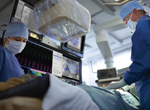
Imaging integration in electrophysiology
EP navigator offers a novel approach to 3D rotational angiography that provides realtime anatomical details of cardiac LA-PV structures with significantly enhanced workflow. For hospital teams working in catheter ablation, capturing the best anatomical detail possible at time of procedure is essential to accurately guide catheters through the anatomy of a beating heart. Philips’EP navigator system provides a novel approach to 3D rotational angiography to capture these images, delivering an alternative to pre-procedural CT/MR imaging to obtain the LA-PV anatomy. Obtaining 3D rotational images can be challenging when treating large patients or performing procedures under general anesthesia. Philips’new EP navigator supports an optimized 3D rotational scan that significantly enhances the workflow for these procedures, by removing barriers encountered in the traditional workflow. In addition, the optimized 3D rotational scan requires less radiation exposure to capture the 3D image, as it uses a shortened trajectory of 159 degrees versus 240 degrees for the traditional scan. To maximize the benefits of advanced X-ray imaging and mapping solutions, integration is critical. Philips and Biosense Webster are collaborating to make such integration a reality. Philips EP navigator already supports seamless integration of the LA-PV anatomy obtained through 3D rotational scans, into Biosense Webster’s CARTO


