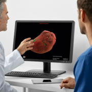
Intelligent software assistant for chest CT
AI-Rad Companion Chest CT is a software assistant that brings artificial intelligence (AI) to computed tomography (CT). Using CT images of the thorax (chest), the software can differentiate between the various structures of the chest, highlight them individually, and mark and measure potential abnormalities. This applies equally to organs such as the heart and lungs, the aorta and the vertebral bodies. The software automatically turns the findings into a quantitative report. AI-Rad Companion Chest CT is the first application based on the new AI-Rad Companion platform. It is designed to help radiologists interpret images faster and more accurately, and to reduce the time involved in documenting results. CT examinations of the thorax are common procedures in daily clinical practice. For radiologists, this means more examinations in a limited amount of time and usually for low reimbursement rates. In radiology, examinations of the chest, a region containing multiple organs, are also challenging because the images display a wide variety of information. Radiologists mainly assess images regarding the primary indication – in other words, the possible disease – which was the reason for performing the CT scan. By contrast, the algorithms in AI-Rad Companion Chest CT pay equal attention to all areas of the chest and can mark abnormalities in places that the radiologist might not consider so closely. The software assistant generates standardized, reproducible, and quantitative reports based on the AI-supported analysis. AI-Rad Companion Chest CT currently supports a variety of tasks, such as identifying lung lesions and calculating cardiovascular risk based on an analysis of coronary artery calcification on non-ECG-triggered CT images. A study in collaboration with the Medical University of South Carolina (MUSC) has also shown that AI-Rad Companion Chest CT can segment and measure the diameter of the aorta, an important parameter for potential aneurysms. AI-Rad Companion Chest CT also examines the spine in the patient’s chest region. It detects and segments the individual vertebrae, labels and analyses them for bone density and possible fractures. This can be helpful for detecting osteoporotic changes at an early stage. AI-Rad Companion Chest CT is a cloud-based solution and uses certified, secure teamplay infrastructure that complies with the Health Information Portability and Accountability Act (HIPAA) in the U.S., and with the General Data Protection Regulation (GDPR) in the EU. The software integrates seamlessly into existing clinical workflows and conforms to Digital Imaging and Communications in Medicine (DICOM) standards. The images and all supporting information can be made automatically available in the picture archiving and communication system (PACS) in line with the radiologist’s individual requirements. The solution is particularly helpful for time-consuming, basic, and repetitive tasks. AI-Rad Companion Chest CT is vendor-neutral and can analyse image data from all CT manufacturers.


