
Scientists discover hidden neural network that may drive early Alzheimer’s disease
Researchers have identified dendritic nanotubes – ultrathin connections between neurons that can transport disease-related proteins. The structures, distinct from conventional synapses, may accelerate toxic protein accumulation in specific brain cells during early Alzheimer’s pathology, offering potential new therapeutic targets.
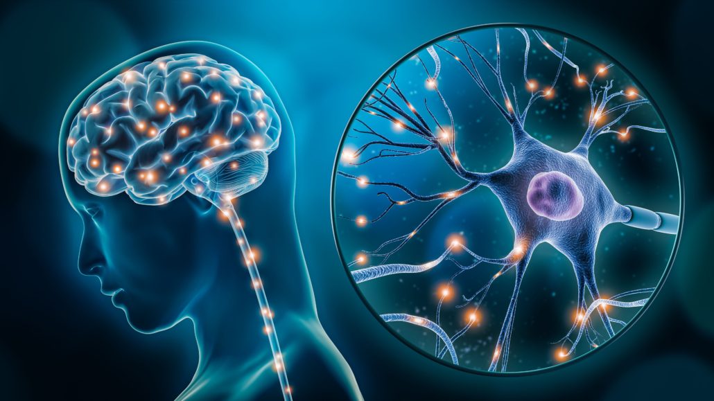
Neuroscientists at Johns Hopkins University have characterised a previously unrecognised communication network in the mammalian brain that operates in parallel with synaptic transmission. The discovery of dendritic nanotubes (DNTs) – membrane bridges approximately 3 micrometres long and just hundreds of nanometres thick – challenges existing understanding of how neurons exchange information and may explain mechanisms underlying neurodegenerative disease progression.
The research team, led by Dr Hyung-Bae Kwon from the Solomon H. Snyder Department of Neuroscience, used advanced super-resolution microscopy combined with machine learning analysis to visualise these structures in both mouse and human brain tissue. Their findings were published in Science on 2 October 2025.
A parallel communication system
“We’ve been looking at the brain forever now, and every once in a while, a surprise comes along,” neuroscientist Lary Walker from Emory University, who was not involved in the work, told Science. Although uncertainties remain about the nanotubes’ basic biology, he suggested the discovery could have wide implications for understanding neuronal communication and disease.
DNTs differ substantially from tunnelling nanotubes previously observed in non-neuronal cells. Whilst traditional tunnelling nanotubes typically exceed 10 micrometres in length with open-ended membrane fusion at terminals, DNTs are shorter structures with closed ends at both contact points. The researchers observed these connections forming directly between dendrites rather than cell bodies.
Using electron microscopy analysis of existing datasets – including the H01 human cortical dataset and mouse somatosensory cortex images – the team documented numerous instances of dendritic filopodia establishing closed-ended membrane contacts with neighbouring dendrites without postsynaptic density. A substantial portion of non-synaptic dendritic filopodia established dendritic contacts, particularly from longer protrusions exceeding 3 micrometres end-to-end distance.
Functional characterisation reveals dual transport mechanisms
The research team demonstrated that DNTs mediate both electrical and molecular communication between neurons. Through calcium uncaging experiments in dissociated cortical cultures, investigators showed that DNTs create pathways for calcium signal propagation between connected neurons. When they artificially increased calcium ion concentration in one neuron, nearby neurons registered corresponding calcium increases – an effect partially blocked by cytochalasin D, which inhibits nanotube formation.
“DNTs actively transported small molecules and human amyloid-β (Aβ),” the authors report in their paper. Using patch-clamp infusion techniques in acute brain slices, researchers injected fluorescently labelled human Aβ1-42 peptides into single layer 5 pyramidal neurons in the medial prefrontal cortex. The peptides spread to neighbouring neurons up to 200 micrometres away, with propagation abolished when nanotube formation was inhibited.
Notably, the research revealed that Aβ transport through DNTs requires active, directional mechanisms rather than passive diffusion. Live-cell imaging captured rapid Aβ transport along neurites at speeds consistent with kinesin or dynein-mediated movement, whilst slower transport along DNT-like extensions matched speeds associated with myosin motor proteins.
Implications for Alzheimer’s disease pathology
The most striking findings emerged from examining APP/PS1 transgenic mice, an established Alzheimer’s disease model. In three-month-old mice – before the formation of extracellular amyloid plaques – the researchers observed significantly elevated DNT formation in the medial prefrontal cortex compared with age-matched controls.
“DNT density increased before plaque formation in the medial prefrontal cortex of APP/PS1 mice,” the authors state, “suggesting that the dendrite-DNT network might play a role in Alzheimer’s disease pathology.”
Computational modelling revealed how altered DNT networks could accelerate disease progression. The simulations predicted that overactivation of the nanotube network drives selective intracellular amyloid accumulation in specific neurons – particularly those with fewer DNT connections. This heterogeneous distribution creates vulnerable cell populations that accumulate toxic protein levels whilst neighbouring neurons maintain lower concentrations.
The research identified a potential pathological feedback mechanism: neurons with high Aβ accumulation showed reduced DNT formation, creating a self-reinforcing cycle. “The toxicity of accumulated Aβ, which impairs cellular metabolism, may further reduce DNTs,” the authors explain. This observation aligns with experimental data showing that whilst mild Aβ exposure (500 nanomolar) increased DNT numbers, higher concentrations (2 micromolar) caused somatic Aβ accumulation and reduced DNT formation.
Methodological advances enable nanoscale tissue imaging
Technical innovations proved crucial for characterising these structures. The team modified super-resolution radial fluctuation (SRRF) microscopy with volumetric deconvolution processing, enabling nanoscale imaging of neuronal structures in cleared brain tissue. This approach, combined with tissue clearing protocols optimised for mouse brain samples, allowed examination of dendritic protrusions at resolutions beyond conventional optical microscopy limits.
Machine learning classification using 22 morphological features distinguished DNTs from synaptic spines with high accuracy. The trained support vector machine models predicted DNT fractions of approximately 12% amongst dendritic protrusions on layer 5 pyramidal neurons in visual cortex – a substantial proportion that had previously escaped detection.
Broader implications for neural connectivity
Beyond Alzheimer’s pathology, DNTs may represent a fundamental component of neural architecture with roles in normal brain function. The structures transmit calcium signals over dendrite-spanning distances within confined regions, potentially supporting local circuit compartmentalisation. The minute-scale dynamics and hours-long lifespans suggest these connections respond rapidly to physiological signals.
“This study provides the first comprehensive characterization of a nanotubular communication network in the brain, establishing a new framework for nonsynaptic signalling between neurons,” the authors conclude. The discovery opens research avenues into brain connectivity, intercellular communication, and mechanisms driving neurological disease.
The findings raise important questions about whether DNTs mediate neuron-glia interactions and transport other disease-associated proteins such as tau. Development of DNT-specific molecular markers would facilitate easier detection and enable investigation of how these networks shape communication across brain cell types.
Reference
Chang, M., Krüssel, S., Parajuli, L. K., et. al. (2025). Intercellular communication in the brain through a dendritic nanotubular network. Science, 390, eadr7403. https://doi.org/10.1126/science.adr7403
