
3D-printed spinal cord scaffolds restore function in paralysed rats
Researchers at the University of Minnesota Twin Cities have demonstrated a groundbreaking process that combines 3D printing, stem cell biology, and lab-grown tissues for spinal cord injury recovery. The innovative approach uses microscopic channels to guide neural regeneration, offering new hope for treating complete spinal cord injuries.
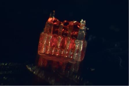
New research combines 3D printing, stem cell biology, and lab-grown tissues for possible treatments of spinal cord injuries. © McAlpine Research Group, University of Minnesota
Novel bioprinting technique creates “mini spinal cords”
The method involves creating a unique 3D-printed framework for lab-grown organs, called an organoid scaffold, with microscopic channels. These channels are then populated with regionally specific spinal neural progenitor cells (sNPCs), which are cells derived from human adult stem cells that have the capacity to divide and differentiate into specific types of mature cells.
The research team, led by Professor Ann Parr from the University of Minnesota’s Department of Neurosurgery, developed scaffolds measuring approximately 1.6mm in width, 0.65mm in height, and 2mm in length. Each scaffold contained three channels with dimensions of approximately 200 micrometers in width and 440 micrometers in height.
“We use the 3D printed channels of the scaffold to direct the growth of the stem cells, which ensures the new nerve fibres grow in the desired way,” said Guebum Han, former University of Minnesota mechanical engineering postdoctoral researcher and first author on the paper. “This method creates a relay system that when placed in the spinal cord bypasses the damaged area.”
Organoid formation demonstrates tissue-specific development
The study, published in Advanced Healthcare Materials, revealed that the 3D-printed constructs developed into functional organoids over time. The authors noted that “3D bioprinted sNPCs in scaffold channels assembled organoids with multiple organ-specific cell types 40 days after printing.”
The researchers observed that cells expressed markers for different neuronal subtypes, including V0, V1, and V2a interneurons, which are crucial for locomotor activity. “V2a spinal interneurons, in particular, are essential for motor control by providing excitatory input to motoneurons,” the authors explained.
RNA sequencing analysis revealed significant differences between 3D-printed scaffold cultures and traditional 2D cell cultures. The 3D constructs showed upregulation of genes associated with organoid formation, regionalization, and post-mitotic neuronal development, indicating enhanced maturation and specialisation.
Functional recovery achieved in complete transection model
The research team tested their approach using a complete spinal cord transection model in rats, where the spinal cord was entirely severed. In their study, the researchers transplanted these scaffolds into rats with spinal cords that were completely severed. The cells successfully differentiated into neurons and extended their nerve fibres in both directions rostral (toward the head) and caudal (toward the tail) – to form new connections with the host’s existing nerve circuits.
Behavioural assessment using the Basso, Beattie, and Bresnahan (BBB) locomotor scale showed significant improvement in rats receiving the organoid scaffolds. At 12 weeks post-transplantation, the organoid scaffold group exhibited a BBB score of 8.4 ± 0.93, compared to 3.6 ± 1.25 for empty scaffold controls and 2.25 ± 0.72 for injury-only animals.
Motor evoked potentials (MEPs), which measure neural connectivity between brain and muscle, were significantly higher in the organoid scaffold group (2.18 ± 0.35 mV) compared to controls (1.07 ± 0.17 mV for empty scaffolds and 0.83 ± 0.18 mV for injury-only).
Integration with host tissue demonstrates therapeutic potential
The new nerve cells integrated seamlessly into the host spinal cord tissue over time, leading to significant functional recovery in the rats. Immunohistochemical analysis at 12 weeks post-transplantation revealed that the majority of transplanted cells differentiated into neurons (63.10 ± 2.31%) and oligodendrocytes (20.21 ± 1.07%).
“Most implanted cells differentiated into neurons and populated both the scaffold channels and the junction between the scaffolds and the host tissue with axonal extension, and the connections were predominantly located in the white matter of the spinal cord sections, both rostral and caudal to the organoid scaffold implants,” the authors reported.
The research demonstrated formation of synaptic connections between transplanted human neurons and host rat neurons, suggesting functional integration rather than mere tissue bridging.
Clinical translation prospects and future directions
“Regenerative medicine has brought about a new era in spinal cord injury research,” said Professor Ann Parr. “Our laboratory is excited to explore the future potential of our ‘mini spinal cords’ for clinical translation.”
The researchers acknowledge that whilst the study represents early-stage research, it offers promising avenues for future clinical applications. “It is envisioned that combining sNPCs, organoid assembly, and 3D printing strategies can ultimately lead to a transformative treatment approach for SCI,” the authors concluded.
The work was funded by the National Institutes of Health, the State of Minnesota Spinal Cord Injury and Traumatic Brain Injury Research Grant Program, and the Spinal Cord Society. The multidisciplinary research team plans to scale up production and continue developing this combination of technologies for future clinical applications.
According to the National Spinal Cord Injury Statistical Center, more than 300,000 people in the United States suffer from spinal cord injuries, yet there remains no proven method to completely reverse the damage and paralysis from such injuries.
Reference
Han, G., Lavoie, N. S., Patil, N., et. al. (2025). 3D-printed scaffolds promote enhanced spinal organoid formation for use in spinal cord injury. Advanced Healthcare Materials, e04817. https://doi.org/10.1002/adhm.202404817
