Pioneering ultrasound research could prevent complications from neck surgery
Researchers in France have embarked on a four-year project to test the new non-invasive technique, that could help surgeons quickly and accurately assess patients for potentially life-changing complications after neck surgery. The research could have global relevance.
Led by surgeons and researchers at world-renowned organisations, including Assistance Publique – Hôpitaux de Paris and Institut Curie, in collaboration with medical device provider Mindray, the research could help to improve safety, outcomes and quality of life for many thousands of neck surgery patients. Physicians believe the programme could also help patients who suffer voice disorders following radiotherapy.
The project aims to reduce reliance on an intrusive procedure known as a nasopharyngoscopy, which involves an endoscope being inserted through the patient’s nose. The procedure, which can require general anaesthetic, is currently used by surgeons in countries around the world, to visualise the larynx as a way to check for complications – in particular vocal cord palsy – after surgery.
A shortage of surgeons and discomfort associated with performing a nasopharyngoscopy has meant that important checks are often not carried out when they should be. This means that complications might not be identified for several months, by which time they are often more serious for patients, and more costly to treat.
Supported by a €500,000 grant from French national research agency ANR, the new research initiative, called VOCALISE, aims to provide an alternative approach, using ultrasound to allow surgeons to see the patient’s larynx after surgery more easily, diagnose any problems before the patient leaves the hospital, and allow for timely treatment and more effective rehabilitation.
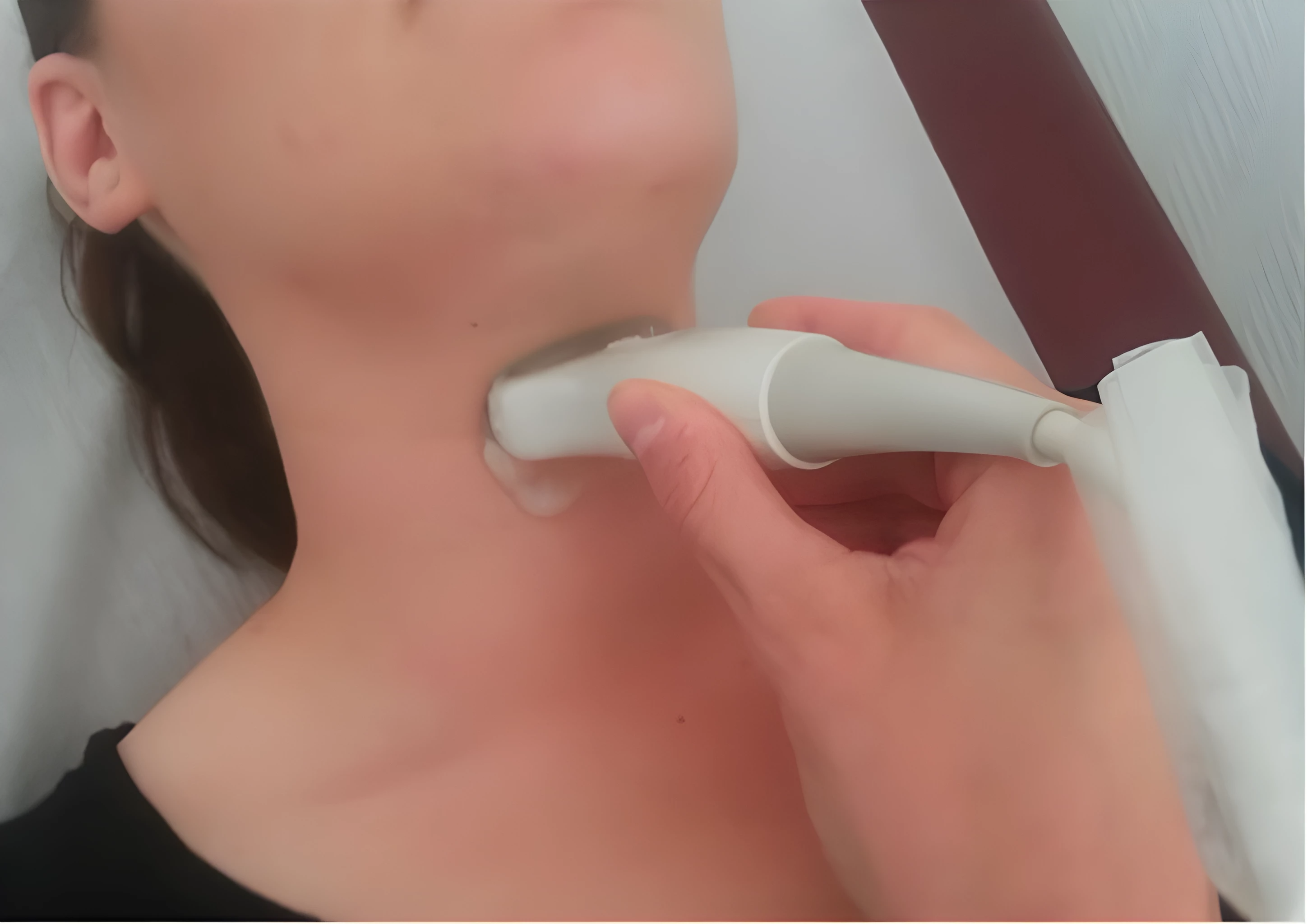
Dynamic TransLaryngeal ultrasound
Known as Dynamic TransLaryngeal ultrasound, a version of the technique has existed for some time, but has so far proven inaccurate for many patients, especially for men, where their laryngeal prominence, or Adam’s apple, can obscure the image, preventing diagnosis.
Researchers believe new ultrasound devices and probes, which have been provided by Mindray, will allow them to overcome these limitations and test their ideas to create a highly accurate and reliable diagnostic approach, that does not expose patients to discomfort or unnecessary risk. Professor Christophe Trésallet, general surgeon for Assistance Publique – Hôpitaux de Paris, said: “Quality of life is a very important part of what we are doing. Vocal fold palsy remains a significant risk for patients undergoing neck surgery, but if we can detect and treat this sooner, then the outcome can be better.
“Specifically designed, intuitive ultrasound devices allow surgeons to do the scan at the patient’s bedside, just after surgery and before they leave hospital. It is easy to use, and junior surgeons are already using it for image acquisition. Within just a few minutes the image can be captured and the patient diagnosed, or further opinion sought when needed.”
Though precise numbers are not known, it is believed that as many as 5% of neck surgery patients develop palsy of the vocal cords. Complications can prevent people being able to work and can include loss of voice, dysphonia, swallowing disorders, and dyspnoea.
Dr Frédérique Frouin, research scientist for Inserm, and permanent researcher at the Laboratory of Translational Imaging in Oncology for Institut Curie, said: “We want to optimise this examination for patients, to make structures visible, and to improve the quality of examination in all cases, but especially for men. Our aim is to show easily if there is a problem, in order to support effective diagnosis, follow up and rehabilitation for patients.
“Our project will measure motion and vibration in the vocal cords. Motion mode within ultrasound, ordinarily seen in other areas such as echocardiography, should provide a better understanding of what needs to be done for the patient. This data will also be complemented with voice recordings to help develop new understanding and correlations.
“Machine learning will also play a role in this initiative, helping to automate parts of the image acquisition and supporting ease of use. Automatic techniques could also have potential to define areas of interest, or even verify if a scan is of an appropriate quality or needs to be repeated.
“Our project could be highly significant for anyone having neck surgery, and we have now identified a second application for radiotherapy patients,” Dr Frouin said.
In total, some 500 participants will take part in the research and information will remain highly secure and confidential.
Partners in the programme include Assistance Publique – Hôpitaux de Paris, Institut Curie, Sorbonne Université, Université Sorbonne Nouvelle, AI developer Apteryx, and Mindray France. Mindray has provided its TE7 devices to support the programme, designed for point-of-care application and ease of use for surgeons. The ultrasound devices have been configured with a specially developed software that allows for semi-automatic calculation of vocal cord motion.
Dr Crystal Lin, the project’s scientific adviser for Mindray, said: “This is an excellent example of leadership in research and medicine – finding new ways to diagnose patients more efficiently and effectively. We are proud to be part of this research team, which is at the cutting edge of using ultrasound for patient benefit. Surgeons are not traditional users of ultrasound but are eager to develop this non-invasive and highly innovative technique. This has the potential to prevent patients presenting to other parts of the busy healthcare system and could make a difference to the lives of patients more widely through Europe and the rest of the world.”
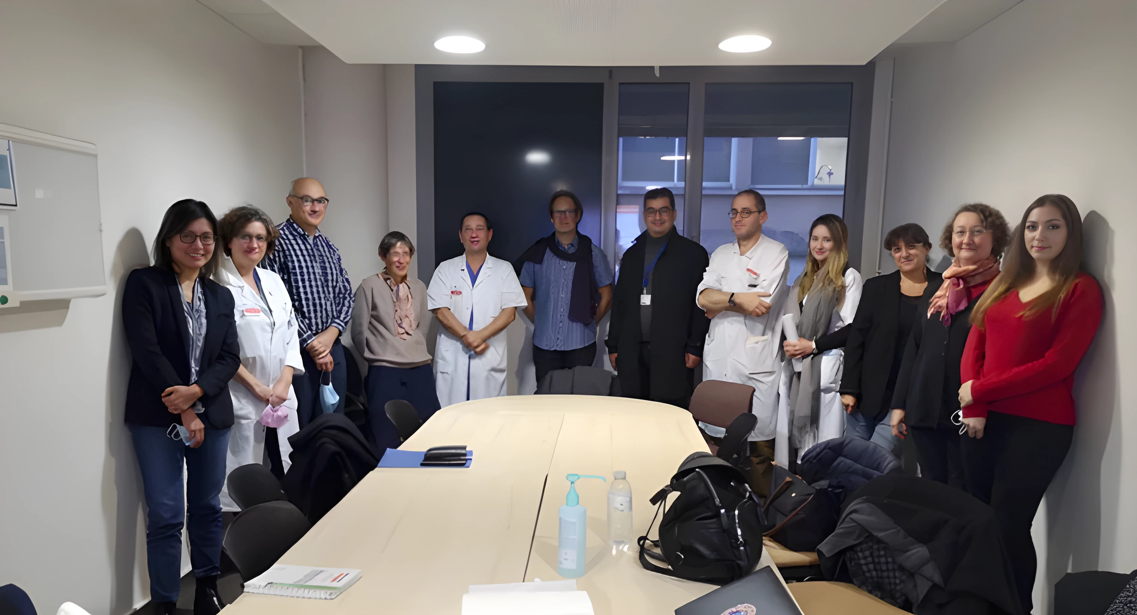
The research team


