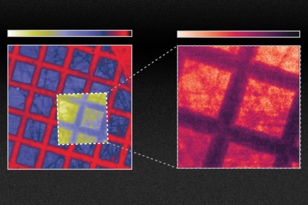
Using nanotech, scientists show how X-ray imaging can be significantly enhanced
Researchers at MIT have shown how the efficiency of scintillators used in X-ray imaging can be improved by at least tenfold, and perhaps even a hundredfold, by changing the material’s surface to create certain nanoscale configurations, such as arrays of wave-like ridges.
Scintillators emit light when bombarded with X-rays. In medical X-ray systems, they convert incoming X-ray radiation into visible light that can then be captured using film or photosensors.
While past attempts to develop more efficient scintillators have focused on finding new materials, the new approach could in principle work with any of the existing materials.
Although it will require more time and effort to integrate their scintillators into existing X-ray machines, the team believes that this method might lead to improvements in medical diagnostic X-rays or CT scans, to reduce dose exposure and improve image quality.
The findings are described in the journal Science, in a paper by MIT doctoral students Charles Roques-Carmes and Nicholas Rivera; MIT professors Marin Soljacic, Steven Johnson, and John Joannopoulos; and 10 others.
Nanotechnology and optical properties
While scintillators have been in use for some 70 years, much of the research in the field has focused on developing new materials that produce brighter or faster light emissions. The new approach instead applies advances in nanotechnology to existing materials. By creating patterns in scintillator materials at a length scale comparable to the wavelengths of the light being emitted, the team found that it was possible to dramatically change the material’s optical properties.
To make what they coined ‘nanophotonic scintillators’, Roques-Carmes says: “You can directly make patterns inside the scintillators, or you can glue on another material that would have holes on the nanoscale. The specifics depend on the exact structure and material.”
For this research, the team took a scintillator and made holes spaced apart by roughly one optical wavelength, or about 500 nanometres.
“The key to what we’re doing is a general theory and framework we have developed,” Rivera says. This allows the researchers to calculate the scintillation levels that would be produced by any arbitrary configuration of nanophotonic structures. The scintillation process itself involves a series of steps, making it complicated to unravel. The framework the team developed involves integrating three different types of physics, Roques-Carmes says. Using this system they have found a good match between their predictions and the results of their subsequent experiments.
The experiments showed a tenfold improvement in emission from the treated scintillator. “So, this is something that might translate into applications for medical imaging, which are optical photon-starved, meaning the conversion of X-rays to optical light limits the image quality. [In medical imaging,] you do not want to irradiate your patients with too much of the X-rays, especially for routine screening, and especially for young patients as well,” Roques-Carmes says.
“We believe that this will open a new field of research in nanophotonics,” he adds. “You can use a lot of the existing work and research that has been done in the field of nanophotonics to improve significantly on existing materials that scintillate.”
Rajiv Gupta, chief of neuroradiology at Massachusetts General Hospital and an associate professor at Harvard Medical School, who was not associated with this work, comments: “The research presented in this paper is hugely significant. Nearly all detectors used in the $100 billion [medical X-ray] industry are indirect detectors,” which is the type of detector the new findings apply to, he says. “Everything that I use in my clinical practice today is based on this principle. This paper improves the efficiency of this process by 10 times. If this claim is even partially true, say the improvement is two times instead of 10 times, it would be transformative for the field!”
Soljacic says that while their experiments proved a tenfold improvement in emission could be achieved in particular systems, by further fine-tuning the design of the nanoscale patterning, “we also show that you can get up to 100 times [improvement] in certain scintillator systems, and we believe we also have a path toward making it even better”.
Nanophotonics
Soljacic points out that in other areas of nanophotonics, a field that deals with how light interacts with materials that are structured at the nanometre scale, the development of computational simulations has enabled rapid, substantial improvements, for example in the development of solar cells and LEDs. The new models this team developed for scintillating materials could facilitate similar leaps in this technology, he says.
Nanophotonics techniques “give you the ultimate power of tailoring and enhancing the behaviour of light,” Soljacic says. “But until now, this promise, this ability to do this with scintillation was unreachable because modelling the scintillation was very challenging. Now, this work for the first time opens up this field of scintillation, fully opens it, for the application of nanophotonics techniques.” More generally, the team believes that the combination of nanophotonic and scintillators might ultimately enable higher resolution, reduced X-ray dose, and energy-resolved X-ray imaging.
This work is “very original and excellent”, says Eli Yablonovitch, a professor of Electrical Engineering and Computer Sciences at the University of California at Berkeley, who was not associated with this research. “New scintillator concepts are very important in medical imaging and in basic research.”
Yablonovitch adds that while the concept still needs to be proven in a practical device, he says: “After years of research on photonic crystals in optical communication and other fields, it’s long overdue that photonic crystals should be applied to scintillators, which are of great practical importance yet have been overlooked” until this work.
References
Charles Roques-Carmes, Nicholas Rivera, Ali Ghorashi, et. al. A framework for scintillation in nanophotonics. Science, 2022; 375 (6583) doi: https://doi.org/10.1126/science.abm9293
MRI innovation makes cancerous tissue light up and easier to see
A new form of magnetic resonance imaging (MRI) that makes cancerous tissue glow in medical images could help doctors more accurately detect and track the progression of cancer over time.
The innovation, developed by researchers at the University of Waterloo, creates images in which cancerous tissue appears to light up compared to healthy tissue, making it easier to see.
“Our studies show this new technology has promising potential to improve cancer screening, prognosis and treatment planning,” said Alexander Wong, Canada Research Chair in Artificial Intelligence and Medical Imaging and a professor of systems design engineering at Waterloo.
Irregular packing of cells leads to differences in the way water molecules move in cancerous tissue compared to healthy tissue. The new technology, called synthetic correlated diffusion imaging, highlights these differences by capturing, synthesizing and mixing MRI signals at different gradient pulse strengths and timings.
In the largest study of its kind, the researchers collaborated with medical experts at the Lunenfeld-Tanenbaum Research Institute, several Toronto hospitals and the Ontario Institute for Cancer Research to apply the technology to a cohort of 200 patients with prostate cancer.
Compared to standard MRI techniques, synthetic correlated diffusion imaging was better at delineating significant cancerous tissue, making it a potentially powerful tool for doctors and radiologists.
“Prostate cancer is the second most common cancer in men worldwide and the most frequently diagnosed cancer among men in more developed countries,” said Wong, also a director of the Vision and Image Processing (VIP) Lab at Waterloo. “That’s why we targeted it first in our research.
“We also have very promising results for breast cancer screening, detection, and treatment planning. This could be a game-changer for many kinds of cancer imaging and clinical decision support.”
Reference:
Wong, A., Gunraj, H., Sivan, V. et al. Synthetic correlated diffusion imaging hyperintensity delineates clinically significant prostate cancer. Scientific Reports 12, 3376 (2022). https://doi.org/10.1038/s41598-022-06872-7


