


Globally HPV is still the most frequent sexually transmitted virus. Certain genotypes cause virtually all cases of cervical cancer, a disease which kills over a quarter of a million women per annum, as well as causing morbidity and mortality from anogenital and oropharyngeal disease in both genders. However back in October 2005 it was reported that Phase III trials, involving twelve thousand women in thirteen countries, had demonstrated that Merck’s quadrivalent HPV vaccine, Gardisil, was 100% effective in preventing pre-malignant cervical lesions. This vaccine, genetically engineered in Brisbane and first licenced for use in public health programmes in Australia, the US, Mexico, Gabon and Europe a decade ago, targets HPV genotypes 6/11 as well as HPV16/18. The former low-risk genotypes cause 90% of anogenital wart infections; it is estimated that the latter high-risk genotypes are responsible for 70% of cervical cancers and 80% to 90% of other HPV-related neoplasms including anal, penile and oropharyngeal cancers. Other vaccines, all of which target the high-risk genotypes HPV 16/18, are now in use. The most recently approved also includes the less common oncogenic genotypes 31/33/45/52/58. HPV vaccine is now approved for use in 129 countries. So after a decade what has been the impact on health from the more than 205 million doses of HPV vaccine that have been distributed worldwide?
The beneficial effect is particularly apparent in countries where there is a high uptake of girls who are vaccinated before they become sexually active. Both infections with HPV and genital warts have plummeted by 90%, with a reduction of 85% in high-grade cervical abnormalities. Data reporting lower numbers of cervical cancer cases post-vaccine will surely follow. The bad news is that the full potential of the vaccine has yet to be realized. Only 64 countries actually include HPV vaccination in their national immunization schedules, and the less developed nations are less likely than the West to have effective programmes that require three timed inoculations and high population coverage. In developed countries such as the US imprudent parents still refuse the vaccine because of possible safety concerns or more bizarrely because they think it will encourage sexual promiscuity in their offspring. However the good news is that in China, which has 28% of the global cervical cancer cases but a particularly cumbersome drug approval process, HPV vaccine has finally been approved and will be available in 2017. Surely a fitting memorial to the late Chinese co-inventor of the initial vaccine, Dr Jian Zhou!

Over the past decade, resuscitation has become one of the fastest growing areas in emergency medical care. The drivers for growth include portable, remote monitoring equipment as well as real time video-consultation. The focus of attention is on cardiopulmonary resuscitation (CPR) and emergency cardiovascular care (ECC), and includes all responses to sudden life-threatening events impacting on the cardiovascular and respiratory system.
Local practices continue to drive growth of best practice in resuscitation. However, there is also substantial cooperation at the global level. The International Liaison Committee on Resuscitation (ILCOR) was founded in 1993 and currently includes representatives from the American Heart Association, the Heart and Stroke Foundation of Canada, the European Resuscitation Council, the Australian and New Zealand Committee on Resuscitation, the Resuscitation Council of Asia, the Resuscitation Council of Southern Africa and the InterAmerican Heart Foundation.
ILCOR members seek to both optimize and minimize international differences in resuscitation practices, but also leave space for geographic, economic, and other real-world differences in practice and the availability of medical devices and drugs.
In 1999, the American Heart Association (AHA) hosted the first ILCOR conference to evaluate best practices and chart resuscitation guidelines. The ILCOR recommendations, formally known as International Consensus on CPR and ECC Science With Treatment Recommendations (CoSTR), were published in 2000. Over the years, ILCOR task forces have evaluated and published CoSTR recommendations in 5-year cycles.
The most recent ILCOR Consensus Conference was held in Dallas in February 2015, and attended by over 230 participants from some 40 countries. Almost two-thirds of participants came from outside the US – giving weight to ILCOR’s position as a global group. The Conference focused, as before, on CPR and ECC, but also covered first aid topics.
One good recent example of the pace of evolution in resuscitation practices is ILCOR’s observation that five years (the task force recommendation cycle) was far too long a period to inform healthcare professionals of therapeutic advances in the field. As a result, it plans to systematically review new science and publish interim advisories on treatment guidelines. The aim is to give resuscitation practitioners access to providing state-of-the-art patient care.
ILCOR’s 2015 CoSTR consensus statements summarize the results of task forces in several areas:
BLS or basic life support (covers quality of CPR and the use of an automated external defibrillator), ALS or advanced life support (post-cardiac arrest care), ACS or acute coronary syndromes, along with education, implementation and teams (EIT), and, for the first time, first aid.
Although dedicated specific task forces cover pediatric BLS and ALS as well as neonatal resuscitation, this review of the 2015 ILCOR guidelines is restricted to adults.
ILCOR task forces perform detailed systematic reviews, evaluate evidence and make recommendations. Task forces identify and prioritize questions using the PICO (population, intervention, comparator, outcome) format, accompanied by a call for public comments. This is followed by a search (with detailed inclusion/exclusion and screening) of relevant articles in three major online databases (PubMed, Embase and the Cochrane Library).
The quality of evidence is tabulated as high, moderate, low, or very low, based on five core domains of risk of bias, inconsistency, indirectness, imprecision, and publication bias (and occasionally other considerations). Together, they follow the so-called GRADE (Grading of Recommendations, Assessment, Development, and Evaluation) methodology for drafting guidelines.
The final wording ranges from ‘we suggest…’ for weak recommendations to ‘we recommend…’ for the strong ones… .’
One of ILCOR’s major goals is continuously-updated and high-quality research into CPR and ECC. An online platform known as SEERS (Scientific Evaluation and Evidence Review System) guides task forces and their individual reviewers, as well as public comments and suggestions. (https://volunteer.heart.org/apps/pico/Pages/default.aspx).
On the other hand, ILCOR also avoids giving attention to areas where there is little development in technology or evidence on practices.
Developments in resuscitation (2010-2015)
The 2015 CoSTR notes that post-OHCA (out-of-hospital cardiac arrest) survival rates are rising, especially when the first monitored rhythm is shockable’ – that is, associated with ventricular fibrillation (VF) or pulseless ventricular tachycardia (pVT). However, survival rates from non-shockable rhythms are also improving. These developments directly correlate with an increased emphasis on improving basic life support (BLS) and advanced life support (ALS).
Given below is a summary of evidence-based recommendations by ILCOR task forces, covering developments since 2010.
Basic life support
EMS dispatchers play the critical role in identifying cardiac arrest, providing CPR instructions to the caller, and activating emergency response. In drowning, it appears that submersion time is a key prognostic factor for outcomes. However, fundamental metrics of high-quality CPR remain the same, with an emphasis on compressions of adequate rate and depth, allowing full chest recoil after each compression, minimizing pauses in compressions, and avoiding excessive ventilation. It is also noted that public access programmes which provide early defibrillation can save many more lives if the programmes are carefully planned and coordinated.
Advanced life support
Post-cardiac arrest care is probably the resuscitation segment undergoing the greatest evolution since 2010, with substantial potential to improve survival from cardiac arrest.
Key recent developments in ALS include results from three major trials on mechanical CPR devices, drug therapy, and insertion of advanced airway devices. In addition, the ALS task force evaluated several studies regarding post-cardiac arrest care and the use of targeted temperature management (TTM).
. Mechanical devices
The three mechanical compression device trials enrolled over 7,500 patients. However, it yielded outcomes similar to those from manual compressions. ILCOR concludes that mechanical CPR devices should not be seen as replacements, but may play a role in conditions where high-quality manual compressions are not feasible.
. Drug therapy
The 2010 CoSTR had pointed to insufficient evidence about drug administration improving survival from cardiac arrest. In 2015, a systematic review identified large observational studies that also challenged routine use of advanced airways and the use of epinephrine for ALS. Since observational studies are known to carry a risk of bias, the findings did not result in a recommendation to change practice. However, they do indicate a need for large randomized controlled trials to assess whether epinephrine and advanced airways are helpful during CPR.
. Targeted Temperature Management
Recent developments in ALS also include greater delineation of the timing and effects of TTM and the need to take account of controlling oxygenation/ventilation and optimizing cardiovascular function. Nevertheless, one high quality TTM trial could not demonstrate an advantage to a temperature goal of either 33C or 36C, while five other trials failed to identify benefits from pre-hospital hypothermia initiation via cold intravenous fluids. Though none of the trials dispelled with the view that post-cardiac arrest patients need a care plan taking account of TTM, there is still little consensus about optimal target temperature and its duration.
Acute coronary syndromes
There are several evidence-based recommendations for ACS since 2010.
. Catheterization, ADP and UFH, troponins
Firstly, pre-hospital ST-segment elevation myocardial infarction (STEMI) activation of a catheterization laboratory treatment delays and improves outcomes.
Secondly, adenosine diphosphate (ADP) receptor antagonists, along with unfractionated heparin (UFH) can be part of a planned percutaneous coronary intervention (PCI) approach and be administered either pre-hospital or in-hospital for suspected STEMI patients. In the pre-hospital setting, enoxaparin is an alternative to UFH. This is not the case with bivalirudin, for which there is insufficient evidence.
Thirdly, the 2015 CoSTR discourages the use of troponins at zero and 2 hours as a standalone measure to exclude ACS diagnosis. Instead, it suggests that negative high-sensitivity troponin I (hs-cTnI) at zero and 2 hours may be used together with low-risk stratification or negative cardiac troponin I (cTnI) or cardiac troponin T (cTnT) measured at zero and 3-6 hours to identify patients at low risk of a major adverse high-sensitivity cardiac troponin I (hs-cTnI) cardiac event (MACE).
. PCI and STEMI
ILCOR’s 2015 CoSTR also has several comments on PCI and STEMI. Its find primary PCI to be generally preferable to fibrinolysis for STEMI reperfusion. However, such decisions must be individualized’ based on time from symptom onset, anticipated delay to PCI, relative contraindications to fibrinolysis, and other patient factors.
Patients with STEMI in the emergency department (ED) of a non-PCI-capable hospital should either be transported rapidly for primary PCI (without fibrinolysis) or be administered fibrinolysis and transported for routine angiography in the first 3-6 hours.
Education, implementation, and teams
One of the most noteworthy areas of attention by ILCOR since 2010 concerns training and continuous quality improvement.
Training cycles
ILCOR states that, although more evidence is needed, it is ‘now recognized’ that training should be more frequent and less time consuming to prevent skill degradation. On the other hand, retraining cycles of 1-2 years are inadequate to maintain competence in resuscitation skills. Though ‘optimal retraining intervals’ remain to be defined, it is clear that more frequent training may help providers likely to encounter a cardiac arrest.
Hi-Fi manikins
ILCOR also suggests replacing standard manikins with high-fidelity manikins at training centres with the infrastructure and resources to maintain the programme.
Performance and quality metrics, social media
Another challenge is that though the role of performance measurement and feedback in cardiac arrest response systems (both in-hospital and out-of-hospital) is recognized, supporting data is of low quality. Closely coupled to improvements in the performance of resuscitation teams is the need for data-driven, performance-focused debriefing.
Finally, ILCOR also notes the rapidly-growing role of social media for notifying suspected OHCA to hospitals and for sourcing bystanders with CPR skills.
First aid
The First Aid Task Force considered stroke assessment, hypoglycemia treatment in diabetics, as well as treatment of open chest wounds and severe bleeding and the identification of concussion.
. Stroke assessment
Observers consider one of the most important recommendations from the First Aid task force is to use stroke assessment systems to improve early identification of possible stroke and enable subsequent referral for definitive treatment. Specific recommendations are made on the FAST (Face, Arm, Speech, Time) tool as well as the Cincinnati Prehospital Stroke Scale, alongside an important observation, that blood glucose measurement could improve the specificity of recognition.
. Hypoglycemia
ILCOR’s 2015 CoSTR observes that first aid providers often face symptoms of hypoglycemia, and a failure to identify and treat it can lead to loss of consciousness and seizures. It recommends administration of glucose tablets for conscious individuals who can swallow, or substitute forms of dietary sugars should glucose tablets not be immediately available.
. Open chest wounds, bleeding, concussion
The 2015 CoSTR recommends that occlusive dressings or devices, or those which might become occlusive, be avoided in the case of open chest wounds in order to avoid engendering a tension pneumothorax.
Recommendations for severe bleeding include using direct pressure, hemostatic dressings and tourniquets – after formal training to ensure effective application and use.
The 2015 First Aid Task Force also recommends developing a simple validated concussion scoring system to accurately identify and manage concussion (minor traumatic brain injury or TBI), which is a condition often encountered by prehospital first-aid providers.
Medicine is in a state of continual progression as new therapies and interventions are developed and technological advances facilitate resuscitation and prolonged organ support. In addition, patients are living longer with increased numbers of comorbidities and complex disease processes. As one result of these demographic changes and medical advances, intensive care units (ICUs) are admitting more patients with a high risk of death, patients who would previously have died before reaching the ICU. As a consequence, the need for end-of-life decisions has become more common than in the past. There has also been a move from an emphasis on survival at all costs to a recognition that the quality of life of survivors must also be taken into account, as well as the quality of dying for those who will not survive.
by Prof Jean-Louis Vincent
With patients who are not considered to have any reasonable chance of benefiting from new or continued intensive care treatments, physicians are faced with four possible options, ranging from continuing with full treatment to support life through to increasing the doses of sedatives to hasten the dying process (Table). In some patients, where withdrawing therapy is permitted, an ICU trial’ can be considered, giving the patient the chance to benefit from a possible intervention. The target of such a test and the time-limit must be set in advance and adhered to; good communication with the family is essential to ensure that these factors are clear. It should be remembered that in some patients, death is actually in their best interest, preventing unnecessary and prolonged suffering.
Recent data suggest that some 40% of ICU non-survivors will have a decision to withhold/withdraw life-sustaining therapy during their ICU stay. Perhaps not surprisingly, there are marked differences in end-of-life decisions and the decision-making practice around the globe. For example, data show that patients are more likely to receive a decision to withhold or withdraw life-sustaining therapy in Oceania, North America, and northern Europe and less commonly in the Middle East, Asia, southern Europe and South America. Although withdrawing and withholding are seen as ethically equivalent in many countries, in others, withholding life-sustaining therapy is considered acceptable but not withdrawing. In Israel, because withdrawal of life-support measures is forbidden, the authorities even passed a law whereby timers can be put on respirators, which then stop by themselves after a preprogrammed time period. The use of sedatives/analgesia at the end-of-life to shorten the dying process also varies considerably among countries and individuals. Some people justify the administration of large doses of sedatives/analgesics in this situation by calling on the double effect’ principle, wherein giving analgesic agents for comfort has the unavoidable effect of hastening death, but this view is rather hypocritical. There is little official guidance available for intensivists regarding this issue and it is perhaps the area of end-of-life management that creates the greatest concern among physicians with fear of possible litigation. The Belgian Society of Intensive Care recently published a statement that ‘Shortening the dying process with use of medication, such as analgesics/sedatives, may sometimes be appropriate, even in the absence of discomfort, and can actually improve the quality of dying’.
The degree of involvement of family members in end-of-life decision making also varies, with families more frequently involved in Northern Europe and the US than in southern European countries. This is in part related to the traditional paternal approach to medical practice still widespread in many southern European countries. Family-centered decision making is also common in East Asian countries, such as Japan, China and South Korea.
The reasons for these international differences are complex. Many are related to the marked cultural and religious diversity among countries. Lack of available resources and financial constraints can also influence end-of-life decision making, particularly in lower income countries. There are also differences among ICUs within a country and among individual intensivists, related again to the cultural and religious backgrounds of the physicians, but also to local legislation, peer and family pressure, and ICU casemix and organization amongst others. The key ethical principles of autonomy, beneficence, non-maleficence and distributive justice must always be used as the basis for any end-of-life decision, but the ways in which these are interpreted and their relative importance may vary according to local factors. It is therefore inappropriate to try and develop a universal consensus on end-of-life decisions as some have suggested, although local guidelines may be useful. Open discussion of these difficult issues must be encouraged within the ICU team and good communication with the family is essential. The aim must always be to provide compassionate end-of-life care, appropriate for the individual patient and his/her particular circumstances.
Suggested reading
Curtis JR and Vincent JL Ethics and end-of-life care for adults in the intensive care unit. Lancet 2010;376:1347-53.
Myburgh J, et al End-of-life care in the intensive care unit: Report from the Task Force of World Federation of Societies of Intensive and Critical Care Medicine. J Crit Care 2016; 34:125-30
Vincent JL, et al ‘Piece’ of mind: end of life in the intensive care unit statement of the Belgian Society of Intensive Care Medicine. J Crit Care 2014;29:174-175
The author
Jean-Louis Vincent, MD, PhD
Dept of Intensive Care,
Erasme University Hospital,
Universite libre de Bruxelles,
Route de Lennik 808,
1070 Brussels,
Belgium
Tel. +32-2-555-3380
Fax +32-2-555-4555
E-mail: jlvincent@intensive.org
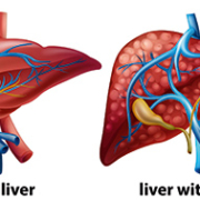
Liver disease is a growing problem across the world. It includes a large range of disorders, such as fatty liver disease (both alcoholic and non-alcoholic), drug-induced liver damage, primary biliary cirrhosis and hepatitis (viral and autoimmune).
Biopsy is gold standard for liver disease
Fibrosis is a relatively common consequence of chronic liver diseases, and its staging, alongside exclusion or confirmation of early compensated cirrhosis, are considered to be vital for surveillance and treatment decisions.
The gold standard for the confirmation of hepatic fibrosis is biopsy. However, biopsy of the liver has several disadvantages. First of all, it is invasive. It is also associated with rare but serious complications. Finally, it can sample only a small portion of the parenchyma (functional rather than connective tissue). This makes it vulnerable to sampling errors.
Non-invasive tests becoming norm
To overcome such constraints, a variety of non-invasive imaging and serological methodologies have been researched and developed for assessing fibrosis. Aside from staging, an ever-growing corpus of data from non-invasive liver tests is also yielding considerable insights for prognostic patient care.
Liver biopsy is now largely restricted to patients showing unexplained discordances in non-invasive testing or those where hepatologists suspect additional etiologies of the disease.
Indeed, non-invasive tests are fast becoming the norm in much of the world, outside the US, although there are several exceptions. The reasons for the lower penetration of non-invasive tests in the US are discussed later.
Ultrasound at forefront
New non-invasive methods for assessing liver fibrosis consists of ultrasound elastography, a diagnostic methodology to evaluate stiffness of tissue, magnetic resonance elastography and serologic testing.
To some of its proponents, elastography is simply a form of the centuries-old systems of diagnosing and assessing diseases via palpation, now extending beyond the scope of physical touch.
While a biopsy is invasive and carries bleeding and infection risks, elastography is seen as a way to get the data needed by clinicians to diagnose and stage liver diseases without the associated complications.
Ultrasound-based elastography is not only used as an alternative to liver biopsy for measuring fibrosis, but also to predict complications in patients with cirrhosis. Another advantage is that elastography, like other non-invasive imaging modalities, can be repeated as often as required to monitor disease progression. Due to their risks, this is simply not feasible with biopsy.
Strain elastography and shear wave elastography
The best-known commercial ultrasound-based techniques for assessing fibrosis include strain elastography and shear wave elastography (SWE). SWE is a real-time two-dimensional elastography technique which enables making quantitative estimates of tissue stiffness in kilopascals (kPa) by virtue of the shear wave speed.
Technologically, even though strain elastography predates SWE, the latter is more easily reproducible than strain elastography, and has rapidly gained interest as the preferred technique. The two are quite different, and outside the hepatology area, seem to have significant complementarities.
Broadly speaking, strain imaging is a qualitative/semi-quantitative method influenced by histotype and lesion size. The use of semi-quantitative indices does not improve performance. Neither does it reduce interoperator variability.
SWE provides accuracy, comparability
Shear wave, on the other hand, is a quantitative method which provides a more accurate and easily comparable assessment of spatial distribution of tissue stiffness.
Most practitioners see SWE as quick and easy to perform, and easily repeated to monitor liver disease progression and measure the effect of a particular treatment. An ultrasound shear wave propagates like ripples of water, as it spreads across tissue. A coherent pattern indicates that a pulse has been applied properly and that there are no artifacts (e.g. from vessels) that would provide erroneous results.
SWE systems provide variable depth of measurement. A depth of 5-6 cms may make it difficult to scan the liver in a large or obese patient, but depths of up to 8 cms are available in certain SWE systems. However, results are not reproducible at such depths, across commercial SWE vendors.
Ease of use not universally accepted
Nevertheless, not everyone agrees that the procedure is easy, especially if SWE results need to be matched against reproducible serological tests. The Society of Radiologists in Ultrasound notes the considerable training required for precision. SWE begins with the positioning of a patient in a left posterior oblique position with the arm raised. Patients need to also breathe slowly, and when asked, suspend breathing, since movement of the liver can reduce accuracy in measurement.
Liver is principal application for SWE
So far, SWE has been used to evaluate and quantify liver fibrosis/cirrhosis of multiple etiologies or with complicating co-morbidities, including chronic hepatitis, liver cancer, steatohepatitis, and biliary atresia. The two-dimensional shear wave elastographic technique offers better performance for assessing liver fibrosis as compared to conventional transient elastography, according to a May 2016 study in the Chinese publication, World Journal of Gastroenterology’.
SWE and hepatitis C
SWE practitioners see it as a tool to assist in earlier detection of conditions such as hepatitis C, and both fatty liver and alcoholic liver disease. Alongside lab studies, SWE offers a means to closely monitor the impact of treatment and assess if the liver will normalize. For many hepatologists, fighting a liver condition before Stage 4 cirrhosis provides a good chance of reversibility.
SWE can also provide information on which hepatitis C patients might benefit from viral therapy. There are numerous reports of patients who would not have been suspected of severe fibrosis or cirrhosis, based on traditional ultrasound grey scaling. At best, the latter provides indicators such as anomalies in the liver contour. However, it does not show signs of cirrhosis such as surface nodularity which are immediately apparent in elastography.
Guiding biopsies
Some clinicians have sought to use SWE to guide liver biopsies and in certain cases, avoid or postpone biopsy. As part of this process, they have addressed one of the major limitations of biopsy, namely restrictions to choice of affected areas, erroneous samples, or inadequacy in sample size enough for interpretation. SWE allows multiple sampling across the liver and generating a mean value. This reduces what in the past would have been a large number of unnecessary biopsies, and minimizes the morbidity of liver biopsy.
SWE in children
SWE has shown specific advantages in pediatric patients. Cincinnati Children’s Hospital Medical Center is gathering data on normal’ stiffness values in children, and on rates of progression, given that published data is almost wholly based on adults.
The study groups cover children with liver transplants, metabolic disorders, cystic fibrosis and those on prolonged intravenous feeding (TPN). One specific area for attention is biliary atresia, a rare but life-threatening condition where the bile ducts in an infant’s liver lack normal openings. The bile builds up and causes damage to the liver.
The pediatric data collection for SWE on newborns with jaundice or cholestasis makes ten measurements. This adds just 5 minutes to a typical ultrasound exam.
Nevertheless, pediatric SWE also has its limitations. According to Dr. Sara O’Hara, who heads the Ultrasound Department at Cincinnati Children’s Hospital, SWE can give variable results in areas such as children with non alcoholic steatohepatitis (NASH) and fatty liver disease.
Breast applications benefit from SWE-plus-strain elastography
In adults, aside from the liver, SWE is seen as a useful technique for evaluation of breast lesions and prostate imaging. In both cases, the technique seems to provide best results in combination with another elastography mode.
For instance, a literature review published in the Journal of Ultrasound’ in 2012 reported that SWE and strain elastography complement each other and overcome mutual limitations in the evaluation of breast lesions.
Clearly, when both types of elastography provide similar results, there is a greater degree of confidence – especially in terms of a near-total elimination of false negatives, which sharply cuts the need for breast biopsies which later prove unnecessary.
There are however some limitations which have been reported in measuring shear wave velocity in the stiffest of breast lesions. Here, rather than propagating through the tumour, the shear wave tends to bounce back. Nevertheless, ongoing improvements in SWE, which have been further reducing examination time and enhancing field of view, means that at some point it could be a tool for breast cancer screening.
Prostate applications benefit from SWE-plus-MR elastography
The use of SWE in prostate cancer, too, shows similar potential for benefits as with breast screening. The first factor is a reduction in biopsies, which prove to have been unnecessary post facto. Studies are under way which seek to correlate stiffness with abnormalities (as well as aggressiveness of tumours) and to assist urologists determine when patients with low-grade prostate cancer must start treatment.
As with SWE and strain elastography in the breast, best results in terms of the prostate are obtained by complementing SWE with another imaging modality – magnetic resonance (MR) elastography. Some findings reveal SWE significantly superior in detecting prostate cancer in the peripheral zone – which is where most tumours occur. However, MR seems to show greater promise in the anterior gland and transitional zone.
Again, as with the breast, the fusion of two modalities permits multiple sampling and tackles a major limitation of prostate biopsy, namely inconvenience and risk, as well as limited choice of affected areas. A few experimental procedures have also targeted fusing MR and SWE images to help guide biopsies.
Using SWE in other organs
SWE has also demonstrated considerable (if still early-stage) promise for evaluating thyroid nodules, indeterminate lymph nodes and uterine fibroids. Another area for investigating SWE include kidney transplants, in order to to avoid excessive biopsies. However, limitations to shear wave captured depth remains a technology challenge for manufacturers to address.
US remains laggard in ultrasound elastography
While most of the world’s regions (Europe, Asia and Latin America) are seeing growth in the use of ultrasound elastography (both SWE and strain), in the US neither is eligible for reimbursement, even in the largest application area – the liver. This is unlike transient elastography, although critics allege it is a blind methodology which neither directly measure fibrosis and often over-estimates it.
Currently, studies in both the US and other parts of the world are seeking to establish the clinical and economic benefits of SWE and strain elastography, including unnecessary invasive biopsies with their associated costs and complications. Eventually, the results of ongoing trials are expected to produce the data which will make ultrasound elastography eligible for reimbursement.
The most self-evident advantage of ultrasound elastography is its non-invasive nature. Unlike a biopsy, it is clearly more feasible to use SWE to screen for patients at greatest risk of chronic liver disease and in need of referral or treatment.

Digital tomosynthesis creates a three-dimensional (3-D) picture using X-rays. In this respect, tomosynthesis is close to a CT (computed tomography) scan. Nevertheless, there are differences between the two. In fact, the development of CT is considered to be one of the reasons for a decline in interest in tomosynthesis, until recently.
One of the principal applications for tomosynthesis today is breast cancer. The basic difference between a digital breast tomosynthesis (DBT) and conventional mammography lies in detail. DBT removes confusing overlying tissue, thus providing clearer imaging and clarity. It also has improved low contrast visibility over mammography, even at a reduced dose. Some explain the difference between DBT and mammography as that of a ball compared to a circle. Nevertheless, in spite of its higher accuracy, the X-ray dose for DBT is similar to that of a mammogram.
Tomosynthesis and CT
Tomosynthesis is now increasingly seen as a low-dose alternative to CT, and is being evaluated against both CT and radiography in several areas, such as erosion in arthritis, or fractures accompanied by metal artifacts.
Technically, tomosynthesis combines digital image capture with the tube/detector motion of CT. However, there are several differences. In CT, the detector makes at least one complete 180-degree (half circle) rotation around the subject. Images are then reconstructed from this data. Tomosynthesis uses a far smaller rotation angle and a lower number of discrete exposures than CT. The lack of comprehensiveness in projections, compared to CT, is compensated by digital processing – with reconstruction of slices at varying depths and thicknesses. The result is that the images are similar to CT, but have a lower depth of field. The reduction in projections, as compared to CT, cuts down on both radiation dosage and cost.
Mammography : the approaches
A mammogram is basically an X-ray examination. However, it uses a machine designed specifically for examining breast tissue. The X-ray format in a mammogram is different while radiation dosages are lower than a conventional X-ray. One of the problems with the latter is that X-rays do not easily penetrate breast tissue. In a mammogram, two glass plates compress the breast to spread out the tissue allowing for a better and more accurate image, using less radiation.
Mammography, formally known as full-field digital mammography (FFDM), usually takes two X-rays of the breast from above (cranial-caudal view, CC) and from an oblique or angled view (mediolateral-oblique, MLO). Single-view mammography uses only the MLO view, and was widespread in the early days of screening. However, it has lower sensitivity and higher recall rates, compared to two-view mammography. Theoretically, the only advantages of single-view mammography are less radiation (which is especially important for young women, who are more sensitive to radiation) and quicker examination speed.
Breast cancer and the mammogram
Breast cancer shows as a typically denser zone than adjacent healthy breast tissue in a mammogram, where it appears as an irregular white area or shadow’.
The term digital’ mammography sometimes confuses patients. However, it simply applies to the storage medium. While regular mammography provides film pictures, digital mammography records images on a computer.
The DBT procedure
While a mammogram is a modified X-ray machine, DBT is delivered by a modified mammogram. It positions the breast in the same way as a mammogram. However, the compression required is less than the latter, in effect just enough for preventing the breast from movement. The X-ray tube then moves around the breast in a circular arc, typically taking 11 X-ray images of 1 mm thickness from different angles, usually over 10 minutes. The images are synthesized by a computer into a clear and highly-focused 3-D image throughout the breast. This allows specialized breast radiologists to see clearly through layers of tissue, including dense tissue, and examine zones of concern from a full range of angles.
Patient comfort is a factor clearly favouring DBT. The breast compression required for a mammogram can be uncomfortable and even sometimes painful, deterring several women from getting tested.
The challenge of false negatives and positives
From a clinical perspective, the high degree of breast compression required by a mammogram can also result in causing folds and overlaps in breast tissue, which can hide the cancer. In other words, negative results do not a guarantee that a woman is cancer-free. The false negative rate is estimated to be as much as 15-20percent. It is also higher in younger women as well as in women with dense breasts.
On the other side, mammograms also face major challenges from false positives. A mammogram may show areas that are considered suspicious or abnormal. This is followed by additional tests (further mammograms, ultrasound and MRI, or an invasive breast biopsy).
One study, published in the May 2014 issue of Annals of Internal Medicine’ found that after 10 years of annual screening mammography, more than half of women will receive at least one false-positive recall.
Some estimates find that 75-80percent of all breast biopsies are unnecessary – that is, they do not find cancers, and 7-9percent receive a false-positive biopsy recommendation. In general, the higher effectiveness of DBT means that patients require fewer (unnecessary) biopsies or other tests.
DBT and multiple tumours
Tomosynthesis also has another major advantage. It has a far greater likelihood than mammography of detecting multiple tumours (which occur in about one in 7 breast cancer patients).
History of DBT
Massachusetts General Hospital (Mass General) in the US is generally credited with pioneering the development and implementation of DBT into a screening programme. In 1992, the hospital’s specialized breast imaging team began researching application of tomosynthesis. In March 2011, just one month after breast tomosynthesis was approved by the US Food and Drug Administration (FDA), Mass General announced that it had performed the first clinical DBT exam in the US. In 2014, the hospital adopted breast tomosynthesis plus mammography as standard protocol for all breast screening.
The hospital states that breast tomosynthesis research in large populations consistently shows ‘improved breast cancer detection rates, especially invasive cancers’ as well as a ‘decrease in call backs, which may lessen anxiety for patients.’
DBT not yet standard of care
Even though digital breast tomosynthesis is now FDA-approved for more than five years, it is not yet considered the standard of care for breast cancer screening. A 2009 recommendation from the US Preventive Services Task Force (USPSTF) has recently been updated. However, it observes that current evidence still remains insufficient to assess the benefits and harms of DBT as a primary screening methodology for breast cancer.
Nevertheless, DBT is available at a small but growing number of US hospitals. These are generally licensed and accredited by the FDA as well as the American College of Radiology (ACR).
DBT in Europe
In Europe, the European Reference Organisation for Quality Assured Breast Screening and Diagnostic Services (EUREF) has recently updated its breast tomosynthesis protocol (version 1.01). Key changes concern technique and methodology (back-projection, dosimetry etc.).
European breast cancer experts frequently cite US studies that show a significant decrease in recall rate using DBT as adjunct to mammography, as well as the increase in cancer detection rates.
Meanwhile, there have recently been several European trials on DBT.
STORM: DBT versus 2D mammograms
The results of one of the first European trials, known as Screening with Tomosynthesis OR standard Mammography (STORM), were published in 2013. This was a prospective comparative study conducted at the University Hospital of Trento, Italy. It sought to determine if DBT overcame some of the limitations of conventional 2D mammography for detection of breast cancer.
The findings were conclusive. The authors of the study estimated that conditional recall could have reduced false positive recalls by 17.2percent without missing any of the cancers detected in the study population.
DBT and mammography combinations studied in Norway
Combinations of DBT with reconstructed 2D images or standard (digital) mammography have also been investigated for screening in Norway.
The Norwegian study was led by a team at Oslo University Hospital, Ullevaal. It sought to compare the performance of two versions of reconstructed two-dimensional (2D) images in combination with DBT versus standard FFDM plus DBT.
Cancer detection rates over two different periods were 8.0 and 7.8 per 1,000 screening examinations for FFDM plus DBT, and 7.4 and 7.7 per 1,000 screenings for reconstructed 2D images plus DBT. False-positive scores were 5.3percent and 4.6percent (over the two periods for FFDM plus DBT, respectively), and 4.6percent and 4.5percent (for reconstructed 2D images plus DBT).
The conclusion of the Norwegian study, published in the June 2014 issue of Radiology’ was clear:
‘The combination of current reconstructed 2D images and DBT performed comparably to FFDM plus DBT and is adequate for routine clinical use when interpreting screening mammograms.’
Sweden: DBT versus mammography, and combinations
Meanwhile, a trial in Sweden, known as the Malmo Breast Tomosynthesis Screening Trial (MBTST), published its results in 2015. MBTST claims to be the first trial designed to assess the efficacy of one-view DBT versus two-view mammography in brast cancer screening, along with a combination of one-view DBT and one-view mammography versus two-view mammography. The authors, from the University of Lund’s Malmo campus found ‘a significant increase in cancer detection rate when using one-view DBT as a stand-alone screening modality, compared to two-view DM (digital mammography). The recall rate increased significantly but was still low.’ They concluded that one-view DBT might be feasible as a stand-alone breast cancer screening modality.
DBT and ultrasound
European researchers have also sought to go beyond comparing DBT with mammography alone. In March 2016, the European CanCer Organisation (ECCO) released interim results from a trial called ASTOUND (Adjunct Screening with Tomosynthesis or Ultrasound in Mammography-negative Dense breasts) at a conference in Amsterdam.
ASTOUND has been recruiting asymptomatic women who attend for breast screening at five imaging centres in Italy and who have extremely dense breasts (defined by the BI-RADS Breast Imaging and Reporting and Data System as being in Categories 3 and 4).
The researchers, led by Dr. Alberto Tagliafico, a radiologist and Assistant Professor of Human Anatomy at the University of Genoa, Italy, have found that adding either DBT or ultrasound scans to standard mammograms could detect breast cancers that would have been missed in women with dense breasts.
Outlook for the future
In general, whether in the US or Europe, more remains to be done to conclusively establish the advantages of DBT in screening. However, it is indisputable that DBT does results in a significant increase in cancer detection rates.
An article in the April 2016 edition of Breast Cancer’ by P. Skane (who led the Norwegian trial mentioned above) argues that ‘DBT should be regarded as a better mammogram that could improve or overcome limitations of the conventional mammography, and tomosynthesis might be considered as the new technique in the next future of breast cancer screening.’
Prins Hendrikstraat 1
5611HH Eindhoven
The Netherlands
info@interhospi.com
PanGlobal Media IS not responsible for any error or omission that might occur in the electronic display of product or company data.
